Connect With Us
Charcot Foot
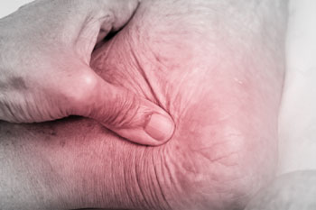
Description
Neuropathic joints, often called Charcot joints, are caused by loss of sensation in the joint so that it is severely damaged and disrupted. The damage and disruption is often so gross that the diagnosis of a neuropathic joint is easily made, both on clinical examination and x-ray, because no one who had sensation would tolerate such destruction of the joint.
Any pathology that leads to loss of sensation in a joint may lead to a Charcot joint.
- Classically, Charcot joints in the lower limb were most often the result of tabes dorsalis but that is very much rarer these days.
- Nowadays the commonest cause is diabetic neuropathy and diabetes is increasing in prevalence.
- In the upper limb the classical cause is syringomyelia.
Diabetic neuropathy is common in developed countries where diabetes is common but in developing countries, tabes dorsalis and leprosy account for a significant amount of neuropathic joints. Charcot joints occur in 10-20% of patients with tabes dorsalis and in 20-25% of patients with syringomyelia.
Presentation
Presentation is variable, depending upon the stage of the disease.
- There may be swelling
- There may be distortion
- There may be loss of function
- Pain is present in 75% but, considering the state of the joint, the clinician will be surprised that there is not considerably more pain.
Treatment
- The patient must be educated about the risk of damaging a joint that is devoid of pain.
- Good control of diabetes is essential to prevent progression of the neuropathy.
- There is a suggestion that biphosphonates may be of value to help heal the bone.
- The affected joint is initially immobilized in a cast. They may still permit ambulation but this should be limited for best results. If there is ulceration the cast must be changed weekly for ulcer evaluation and debridement. Plain x-rays every month help evaluate progress. The cast is usually on for 3 to 6 months.
- After removing the cast, protection of the joint for the rest of the patient’s life is essential. This requires patient education and foot care by a podiatrist. Various types of braces may protect the foot.
- Various types of protective shoes may be required. If ulcers are present, a rocker-bottom sole can be used. A less intense regime may be permitted after 6-24 months, depending upon clinical progress but special footwear is required for life.
- The total process of healing usually takes 1-2 years. Preventing further injury, noting temperature changes, checking feet every day, reporting trauma, and receiving professional footcare also are important aspects of treatment.
- If conservative treatment fails, surgical options will be considered.

